Scimitar ear drum, a rare congenital heart defect, is characterized by an anomalous blood vessel that connects the inferior vena cava to the right atrium. This unique vessel resembles a scimitar, a curved sword, and gives the condition its name.
Scimitar ear drum can cause a range of symptoms, from mild to severe, and requires specialized medical attention for proper diagnosis and management.
This condition, often diagnosed in infancy or childhood, can have a significant impact on the cardiovascular and pulmonary systems. Understanding the causes, symptoms, and treatment options for scimitar ear drum is crucial for healthcare professionals and individuals affected by this condition.
Scimitar Syndrome

Scimitar Syndrome is a rare congenital heart defect that affects the pulmonary veins and the right lung. It is characterized by the presence of a partial or complete anomalous pulmonary venous return from the right lung, which means that the pulmonary veins from the right lung do not connect to the left atrium as they should, but instead connect to the inferior vena cava.
This abnormal connection can lead to a number of problems, including: decreased blood flow to the right lung, increased blood flow to the left lung, and pulmonary hypertension (high blood pressure in the lungs).
Signs and Symptoms
The signs and symptoms of Scimitar Syndrome can vary depending on the severity of the defect. Some people with Scimitar Syndrome may have no symptoms at all, while others may experience a range of symptoms, including:
- Shortness of breath
- Fatigue
- Chest pain
- Wheezing
- Cyanosis (bluish tint to the skin, lips, or nail beds)
- Recurrent respiratory infections
Prevalence and Demographics
Scimitar Syndrome is a rare condition, affecting approximately 1 in 100,000 people. It is more common in females than males, and it is often associated with other congenital heart defects, such as tetralogy of Fallot and truncus arteriosus.
Clinical Manifestations

Scimitar Syndrome is a rare congenital heart defect that affects the development of the heart and lungs. The clinical manifestations of Scimitar Syndrome can vary depending on the severity of the defect. However, there are some common cardiovascular and pulmonary manifestations associated with this condition.
Cardiovascular Manifestations, Scimitar ear drum
- Pulmonary hypertension: This is a condition in which the blood pressure in the arteries of the lungs is abnormally high. Pulmonary hypertension can lead to shortness of breath, fatigue, and chest pain.
- Right-sided heart failure: This is a condition in which the right side of the heart is unable to pump blood effectively. Right-sided heart failure can lead to swelling in the legs, ankles, and feet, as well as shortness of breath and fatigue.
- Atrial septal defect (ASD): This is a hole in the wall between the two upper chambers of the heart. An ASD can allow blood to flow from the left atrium to the right atrium, which can lead to pulmonary hypertension.
- Ventricular septal defect (VSD): This is a hole in the wall between the two lower chambers of the heart. A VSD can allow blood to flow from the left ventricle to the right ventricle, which can lead to pulmonary hypertension.
Pulmonary Manifestations
- Hypoplastic lung: This is a condition in which one lung is underdeveloped. Hypoplastic lung can lead to shortness of breath, fatigue, and chest pain.
- Pulmonary artery sling: This is a condition in which the pulmonary artery wraps around the trachea and esophagus. Pulmonary artery sling can lead to shortness of breath, difficulty swallowing, and chest pain.
- Tracheomalacia: This is a condition in which the trachea is weakened and collapses. Tracheomalacia can lead to shortness of breath, wheezing, and coughing.
Extra-pulmonary Manifestations
- Gastrointestinal problems: Some people with Scimitar Syndrome may experience gastrointestinal problems, such as reflux, constipation, and diarrhea.
- Neurological problems: Some people with Scimitar Syndrome may experience neurological problems, such as seizures, developmental delays, and learning disabilities.
- Skeletal problems: Some people with Scimitar Syndrome may experience skeletal problems, such as scoliosis and kyphosis.
Diagnostic Evaluation

Scimitar Syndrome requires comprehensive evaluation to confirm the diagnosis. Imaging techniques, such as chest X-ray, echocardiography, and cardiac catheterization, play crucial roles in assessing the anatomical abnormalities and physiological consequences of the condition.
Chest X-ray
Chest X-ray provides an initial screening tool to detect the characteristic findings of Scimitar Syndrome. The X-ray may reveal an abnormal pulmonary artery, known as the scimitar vein, which is smaller than the usual pulmonary arteries. Additionally, it can show enlargement of the right atrium and right ventricle, as well as dextroposition of the heart, indicating a shift of the heart to the right side of the chest.
Echocardiography
Echocardiography is a non-invasive imaging technique that uses sound waves to visualize the heart’s structure and function. It is particularly useful in evaluating Scimitar Syndrome, as it can provide detailed images of the heart chambers, valves, and blood flow patterns.
Echocardiography can confirm the presence of the scimitar vein, assess the severity of pulmonary artery stenosis, and evaluate the function of the right ventricle.
Cardiac Catheterization
Cardiac catheterization is an invasive procedure that involves inserting a thin tube (catheter) into the heart through a blood vessel. It is used to confirm the diagnosis of Scimitar Syndrome by directly measuring the pressures within the heart chambers and pulmonary arteries.
Cardiac catheterization can also be used to perform balloon angioplasty or stent placement to widen narrowed pulmonary arteries and improve blood flow.
Management and Treatment
:max_bytes(150000):strip_icc()/what-causes-a-retracted-ear-drum-1191976-5c04ac1e46e0fb0001dd5eba.png)
Scimitar Syndrome management encompasses both surgical and non-surgical approaches. Surgical intervention is indicated in severe cases, while non-surgical measures provide supportive care and address associated conditions.
Surgical Management
Surgical management of Scimitar Syndrome aims to correct the anomalous pulmonary venous drainage and address associated cardiac defects. The specific surgical approach depends on the individual patient’s anatomy and the severity of the condition.
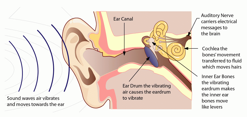
Scimitar ear drum is a rare congenital malformation of the middle ear. It’s characterized by an abnormally shaped eardrum that resembles a scimitar, a curved sword. If you’re interested in learning more about the history and significance of scimitar-shaped objects, you can check out scimitar drums.
Scimitar ear drum can cause hearing loss and other problems, so it’s important to see a doctor if you suspect you have it.
- Anomalous Pulmonary Venous Drainage Correction:This surgery involves redirecting the anomalous pulmonary veins to drain directly into the left atrium, bypassing the IVC.
- Cardiac Defect Repair:If present, associated cardiac defects, such as atrial septal defect (ASD) or patent ductus arteriosus (PDA), are repaired during the same surgery.
Surgical outcomes are generally favorable, with significant improvement in symptoms and long-term survival. However, the complexity of the surgery and the patient’s overall health status influence the outcomes.
Non-Surgical Management
Non-surgical management focuses on providing supportive care and addressing associated conditions without surgical intervention. This approach is typically recommended for patients with mild to moderate Scimitar Syndrome who are asymptomatic or have minimal symptoms.
- Medical Management:Medications, such as diuretics and ACE inhibitors, may be prescribed to manage fluid overload and improve cardiac function.
- Respiratory Support:Oxygen therapy or mechanical ventilation may be necessary in severe cases with respiratory distress.
- Pulmonary Hypertension Management:If pulmonary hypertension develops, specific medications or procedures may be used to lower pulmonary artery pressure.
- Regular Monitoring:Patients with Scimitar Syndrome require regular follow-up with a cardiologist to monitor their condition and adjust treatment as needed.
Non-surgical management aims to improve symptoms, prevent complications, and optimize the patient’s quality of life.
Differential Diagnosis
The differential diagnosis of Scimitar Syndrome involves considering conditions that can mimic its symptoms, including:
Conditions Mimicking Scimitar Syndrome
- Pulmonary atresia with intact ventricular septum (PA/IVS)
- Total anomalous pulmonary venous return (TAPVR)
- Tricuspid atresia
- Ebstein’s anomaly
- Congenital diaphragmatic hernia (CDH)
- Pleural effusion
- Pneumonia
Differentiating Features
The key differentiating features between Scimitar Syndrome and its mimics are summarized in the table below:
| Feature | Scimitar Syndrome | Mimics |
|---|---|---|
| Anomalous pulmonary venous return | Anomalous right pulmonary vein drains into the inferior vena cava | Varies depending on the specific mimic |
| Cardiac defects | Usually present, such as right ventricular outflow tract obstruction or pulmonary atresia | May or may not be present |
| Pleural effusion | Often present on the right side | May or may not be present |
| Lung hypoplasia | May be present in the right lung | May or may not be present |
| Other anomalies | May include skeletal, renal, or gastrointestinal anomalies | Varies depending on the specific mimic |
Epidemiology and Etiology

Scimitar Syndrome is a rare congenital heart defect with an estimated prevalence of 1 in 100,000 live births. It is more common in females than males, with a ratio of 2:1.The embryologic basis of Scimitar Syndrome involves abnormal development of the right pulmonary artery and lung during fetal development.
The right pulmonary artery, which normally arises from the main pulmonary artery, fails to develop properly and instead arises from the inferior vena cava. This abnormal connection leads to a reduction in blood flow to the right lung, resulting in the characteristic scimitar-shaped appearance of the right lung on chest X-ray.Genetic factors are also believed to play a role in the development of Scimitar Syndrome.
Mutations in the genes encoding for proteins involved in the development of the heart and lungs have been linked to the condition. However, the exact genetic basis of Scimitar Syndrome is still not fully understood.
Prognosis and Outcomes: Scimitar Ear Drum
Scimitar Syndrome has a variable prognosis, influenced by several factors. Early diagnosis and intervention are crucial for improving outcomes.
Factors Influencing Prognosis
- Severity of cardiac anomalies:More severe heart defects, such as pulmonary atresia, result in a poorer prognosis.
- Associated anomalies:Other associated conditions, such as Down syndrome or tracheoesophageal fistula, can impact the overall prognosis.
- Timely intervention:Early surgical correction of cardiac defects can significantly improve outcomes.
- Patient’s age at diagnosis:Infants and younger children have a better prognosis than older children and adults.
Long-Term Outcomes
With appropriate medical management and surgical interventions, many individuals with Scimitar Syndrome can lead full and active lives. Long-term outcomes include:
- Improved cardiac function:Surgery can restore normal blood flow and heart function.
- Reduced risk of complications:Early intervention can prevent serious complications, such as pulmonary hypertension and heart failure.
- Normal life expectancy:With proper care, most patients with Scimitar Syndrome can have a normal life expectancy.
Imaging Findings

Scimitar Syndrome is characterized by a constellation of anomalies affecting the heart and lungs, and imaging plays a crucial role in confirming the diagnosis and assessing its severity.
Chest X-ray Findings
Chest X-ray findings in Scimitar Syndrome typically include:
- Prominent pulmonary artery extending from the inferior vena cava towards the right lung.
- Hypoplasia or absence of the right pulmonary artery.
- Cardiomegaly due to right ventricular enlargement.
- Deviation of the trachea and esophagus to the left due to mediastinal shift.
Echocardiographic Findings
Echocardiography provides detailed images of the heart and great vessels, and can reveal the following findings in Scimitar Syndrome:
- Abnormal connection of the inferior vena cava to the right atrium.
- Hypoplasia or absence of the right pulmonary artery.
- Right ventricular enlargement and hypertrophy.
- Increased pulmonary blood flow to the right lung.
- Atrial and ventricular septal defects in some cases.
Computed Tomography (CT) and Magnetic Resonance Imaging (MRI)
CT and MRI provide cross-sectional images of the chest, allowing for detailed evaluation of the heart, lungs, and mediastinum. These modalities can further confirm the presence of anomalous pulmonary veins, assess the degree of pulmonary artery hypoplasia, and visualize any associated anomalies, such as diaphragmatic eventration or pericardial effusion.
Historical Perspective
Scimitar Syndrome, initially described in 1958 by Elliott and Schiebler, gained recognition in 1960 when Neill and associates coined the term “scimitar syndrome.” Early descriptions focused on the characteristic pulmonary artery anomaly and the associated cardiac and vascular defects.
Over the years, diagnostic modalities have evolved significantly, enabling more precise identification of anatomical variations and functional abnormalities. Advancements in surgical techniques and perioperative care have improved patient outcomes, contributing to a better understanding of the natural history and prognosis of Scimitar Syndrome.
Current Understanding and Future Directions
Current research aims to further elucidate the genetic basis, embryological development, and long-term outcomes of Scimitar Syndrome. Studies are exploring the role of genetic factors, environmental influences, and epigenetic modifications in the pathogenesis of the syndrome.
Ongoing investigations focus on identifying biomarkers for early diagnosis, developing personalized treatment strategies based on individual patient characteristics, and evaluating the impact of interventions on long-term outcomes. Advances in imaging techniques, such as 4D flow magnetic resonance imaging, offer promising insights into hemodynamic changes and provide valuable information for surgical planning.
Answers to Common Questions
What are the common symptoms of scimitar ear drum?
Symptoms can vary depending on the severity of the condition, but may include shortness of breath, fatigue, chest pain, and cyanosis (bluish tint to the skin).
How is scimitar ear drum diagnosed?
Diagnosis typically involves a combination of physical examination, chest X-ray, echocardiography, and cardiac catheterization.
What are the treatment options for scimitar ear drum?
Treatment options vary depending on the individual’s condition and may include surgical intervention, medication, or a combination of both.